Letter to the Editor: “Optimizing resources in radiology practice amidst COVID-19 pandemic: preliminary experience from a tertiary care hospital in India”
by Abanti Das, Rohini Gupta Ghasi, Ritu Nair Misra (das_abanti@yahoo.co.in)
India is currently poised precariously at the growing curve of the globally emerging pandemic of corona virus disease (COVID-19). With more than 1000 new confirmed cases being added daily, the tally of total cases stands close to a staggering 30,000 as on 26th April 2020. Globally, as the toll of infected patients inches close to three million, India is gradually scaling up the list of affected countries worldwide and is currently positioned 16th globally and 4th in Asia in terms of total affected patients and total deaths, following close on the heels of China [1].
This rapidly unfolding pandemic has posed peculiar and novel challenges to the healthcare system which is struggling under the exponential rise of infection worldwide, more so in developing nations like India with relative paucity of health care resources. Radiology has come to play an important complementary role in the concerted effort to overcome the pandemic and majority of the upcoming COVID-19 literature on radiology is focused on the role of chest radiography and CT in screening, diagnosis and follow-up of patients [2-4].
Various institutions have proposed their radiology preparedness in dealing with the work flow of patients in current scenario [5, 6]. However, adequate infection control issues related to transport of COVID-19 positive patients to CT suite, proper decontamination of CT room and attendant downtime, lack of availability of sufficient number of CT scanners in various parts of the world, mobile chest radiography is widely being used for diagnostic and prognostic purposes in these patients [7].
Most of the guidelines and consensus statements refer to workflow and infrastructure of radiology departments in developed nations with access to adequate number and quality of CT scanners and digital radiography units. The scenario may, however, not be as streamlined in developing nations like India where limited availability of CT scanners and digital radiography units in hospital catering to far-flung areas is a common problem. Hence, meticulous observation of these aforementioned guidelines may not always be feasible. There has been a lot of emphasis on use of portable radiography in the current pandemic scenario to strike a balance between reduced patient mobility and chances of in-hospital cross-contamination as well as adequate diagnostic imaging.
Most of the articles mention use of portable digital radiography units for this purpose, which are faster, less cumbersome and involve minimal patient contact with the radiographer. However, in certain resource-poor areas, portable computed radiography (CR) radiography units are still the most commonly used and often, the only available mobile radiography unit in the hospital. Imaging COVID-19 positive patients using CR radiography units pose unique challenges in terms of ensuring adequate protection of the radiographer as well as of other patients. We, in this correspondence wish to share our experience from a large 2500 bedded tertiary care public hospital with designated COVID-19 facility in New Delhi, India.
Two dedicated portable CR radiography units are deployed for our COVID-19 designated ward and ICU both of which are stationed in a separate building away from the main hospital area. One CR unit is kept in reserve in case of any technical issue or breakdown. In order to ensure adequate protection of the radiographers as well as to maintain a steady workflow in COVID-19 designated ward , we devised an indigenous algorithm for the same. The steps are enumerated as follows:
1. After a call is received for portable chest radiograph of a COVID-19 positive or suspected patient, the CR cassette, which is to be used, is first wrapped up in two plastic sheets and taped securely at the back to ensure complete coverage. We use polythene garbage disposal bags for the same which are readily available in our hospital supply. Twelve CR cassettes, two of size 10”x10” (pediatric patients), five each of 14”x14” (for adults) and five each of 17”x14” (for adults) are reserved for COVID ward and ICU (Fig.1).
2. For each round of portable radiographs, the radiographer is accompanied by an assistant to the ward and ICU. Radiographer enters the ward in level 3 protection complete with sealed personal protective equipment, double latex gloves, goggles, N95 mask, face shield, head cap and shoe-cover. The double-bagged plastic enclosed cassette is then positioned underneath the patient for frontal chest radiography. The assistant meanwhile waits outside the ward.
3. After exposure, the outer plastic cover is discarded in the ward and disposed of as per guidelines for biomedical waste management. The CR cassette enclosed in inner plastic cover is disinfected with 95% alcohol containing solution and handed over to the assistant waiting outside the ward who brings it back to the radiology unit where the CR reader is positioned.
4. The inner plastic cover is removed in radiology unit and the cassette is wiped clean with 95% alcohol containing solution including all the surface and edges (Fig.2).
5. After allowing it to dry up, the cassette is inserted into the slot of laser reader to allow for image processing. The image thus obtained contains no or minimal artifacts due to the overlapping folds of external plastic cover but they are not significant enough to hamper image interpretation (Fig.2).
6. The portable CR radiography unit is also similarly double-coated with polythene garbage disposal bag and after every round of ward and ICU, the outer sheet is disposed of in the ward and the machine is parked in a secure space of the ward (Fig.3).
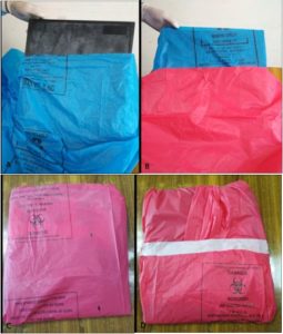
Fig1. (A,B) Double-bagging of CR cassette in polythene garbage disposal bags (inner:blue, outer:pink). (C) Front and (D) back of securely taped double-bagged CR cassette.

Fig2. (A) Exposed CR cassette as received in radiology unit with outer (pink) polythene garbage bag removed and wrapped in disinfected inner polythene (blue) garbage bag. (B) Second round of disinfection of cassette surfaces with 95% alchohol solution. (C) Insertion in CR cassette reader for image processing. (D) Final chest radiograph with no artifacts.
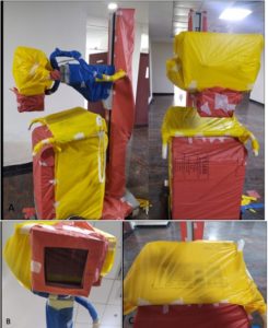
Fig.3 (A, B, C) Portable CR radiography unit double-wrapped in polythene garbage disposal bags with special cut-outs for X-ray imaging window for COVID-19 patients.
The entire process requires approximately 20-25 minutes per patient, but with the availability of two portable CR units and 10 cassettes, we are currently able to manage the patient throughput and can ramp up our capacity in case of patient surge. We have performed 264 portable chest radiographs since 20th February 2020 till 26th April 2020 with an average of about 4 cases per day following this drill. There has not been a single report of cross-infection of any of our radiology technical staff or radiographer posted on COVID-19 duty during this period.
Image acquisition and processing using portable CR radiography unit is, understandably, a tedious and time-consuming process and may sometimes affect patient throughput in a busy radiology unit. Working with this system also entails longer and more frequent contact with cassette surface which comes in contact with the patient. This surely source of anxiety for the radiographers and technical staff as for all the health care workers who are working in close contact with COVID-19 positive patients.
We have tried to incorporate the guidelines and recommendations as issued from time to time by various regulatory bodies, both in India and outside regarding reorganization of available resources and temporary suspension of non-urgent and non-emergent procedures to redirect more of the work force for COVID-19 preparedness. However, as pointed out earlier, most of these recommendations are proposed by institutions and centers with an ideal radiology set-up and with availability of state-of-the art equipment. Resource-constraint nations like India with most of the hospitals in rural and remote areas lacking adequate number and quality of radiology equipment are struggling to cope with the burgeoning case load of COVID-19. Hence, we need to devise our own indigenous algorithms to manage whatever available resources we have so that they can be best utilized in these times of crisis.